Most unusual medical stories get swept into the world of internet mystery or sensational headlines, but this one caught my attention for a very different reason. It’s not true crime, not paranormal, and not science fiction—just an astonishing look at how complex human development can be. The case involves a child whose delayed motor development and enlarged head circumference led doctors to a diagnosis so rare that fewer than 20 similar cases have ever been reported.
An Unusual Diagnosis
When the child was brought in for medical evaluation, imaging revealed hydrocephalus, an abnormal buildup of fluid inside the brain’s ventricles. This pressure was compressing surrounding brain tissue, so doctors performed additional scans to understand the source of the problem. What they found was remarkable: inside the brain’s ventricular system was a mass containing bone-like structures. Further imaging showed the presence of a vertebral column and long bones—features resembling a malformed fetus.
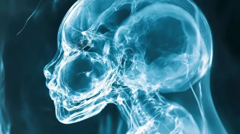
This led doctors to diagnose an exceptionally rare condition known as fetus in fetu occurring inside the skull. While the discovery was startling, doctors emphasized that this was not supernatural or fantastical. Instead, it represented a rare developmental anomaly rooted in the earliest stages of embryonic growth.
What Is Fetus in Fetu?
Fetus in fetu is an extremely rare congenital condition in which a malformed twin becomes enclosed within the body of its sibling during development. It is considered a form of abnormal twinning involving genetically related embryos. The condition occurs in roughly 1 in 500,000 live births. Most cases involve abdominal masses, often located behind the abdominal organs in the retroperitoneal space.
Intracranial cases, however—where the malformed twin-like structure appears inside the skull—are exceedingly rare. Fewer than 20 such instances have ever been documented in medical literature. That rarity is part of what made this case so scientifically intriguing.
What Doctors Found Inside the Skull
Imaging revealed that the mass inside the child’s brain included a vertebral column, femur, and tibia. There were signs consistent with spina bifida, indicating incomplete closure of the malformed structure’s spine. After surgical removal, doctors observed additional limb-like components, including upper limbs and finger-like buds.
Genetic testing provided further confirmation: both the child and the mass shared identical single-nucleotide variants. This strongly supported the conclusion that the mass represented a malformed twin rather than a tumor or unrelated growth.
Researchers proposed that the anomaly may have resulted from unseparated blastocysts early in embryonic development. During the folding of the neural plate, part of one embryo may have been enveloped within the forming forebrain of the other.
How This Differs From a Tumor
Conditions like fetus in fetu can resemble certain tumors, especially teratomas, which can contain mixed tissue types. However, the key distinguishing factor is organization. Fetus in fetu often contains recognizable body-axis structures—such as a vertebral column or limb buds—whereas teratomas tend to be disorganized masses.
This distinction is medically important because teratomas can behave differently, carry their own risks, and require specific forms of follow-up. In this case, the presence of organized fetal-like structures and the genetic evidence confirmed the diagnosis of intraventricular fetus in fetu.
The Surgery and What Happened After
Doctors removed the mass surgically because it was causing dangerous pressure and fluid buildup in the child’s brain. The structure measured roughly 10 centimeters—about four inches long. The original medical report detailed the diagnosis and the surgical removal but did not provide long-term follow-up information. Because of that, it’s important not to assume anything about the child’s outcome beyond what was written.
Even without long-term data, the case itself remains vitally important to medical science. It helps researchers better understand rare developmental anomalies and highlights how modern imaging, surgical techniques, and genetic testing can illuminate conditions that would once have been totally mysterious.
A Rare Window Into Early Development
This case is not evidence of a conscious twin living inside the brain. The malformed structure was not viable and had no neurological function. Instead, it was the result of an extraordinarily rare developmental error early in embryonic life.
Still, it is deeply fascinating. It offers a rare glimpse into how intricate—and occasionally imperfect—the earliest moments of human development can be. For physicians and researchers, the lesson is scientific, not sensational: congenital anomalies can appear in places no one expects, and careful analysis is essential for understanding them.
Cases like this remind us that the human body, even from its very first moments, is full of complexity, surprise, and the occasional phenomenon that challenges even seasoned medical experts.
 amandasheppardauthor